Investigations
Your Organisational Guidance
ebpracticenet urges you to prioritise the following organisational guidance:
Richtlijn acute rinosinusitisPublished by: Werkgroep Ontwikkeling Richtlijnen Eerste Lijn (Worel)Last published: 2023Guide de pratique clinique rhinosinusite aiguëPublished by: Groupe de travail Développement de recommandations de première ligneLast published: 20231st investigations to order
clinical diagnosis
Test
Laboratory tests and imaging studies are not indicated for evaluation of routine, uncomplicated acute rhinosinusitis. Imaging may be warranted in the case of complications, recurrent episodes of rhinosinusitis, suspected anatomical abnormalities, rapidly progressing or suspected acute invasive fungal rhinosinusitis, or if an alternative diagnosis is suspected.[24]
Result
diagnosis is based on history and physical examination
Investigations to consider
nasal endoscopy
Test
Recommended in selected patients (e.g., patient refractory to empirical antibiotic therapy, concern for antibiotic resistance, patient immunocompromised) as it can provide excellent visualisation of the nasal cavity and sinuses.
There are two types of endoscope: rigid and flexible.
Rigid: has superior resolution and only requires the use of one hand. This easily allows cultures of the nasal cavity or sinus to be obtained if necessary.
Flexible: more comfortable for patients, but requires both hands to use. It is preferred in children as it is better tolerated.
Either type may be used in adults and children. Choice will depend on the practitioner's familiarity with the procedure, and most will be performed by an ear, nose, and throat specialist.
[Figure caption and citation for the preceding image starts]: Nasal endoscopy of the left nasal cavity showing a small polyp and pus in the middle meatusFrom the collection of Joseph K. Han [Citation ends].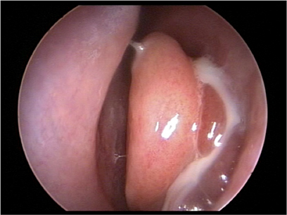
Result
mucosal erythema, purulent discharge
sinus culture
Test
Not required for diagnosis but can be helpful for planning the appropriate management plan for the patient, especially if the patient is refractory to empirical antibiotic therapy, if there is a concern for antibiotic resistance, or if the patient is immunocompromised.
Endoscopic culture taken from the sinus drainage path is well tolerated, and has a high correlation with the pathogenic bacteria, especially when there is purulence in the middle meatus; it is therefore the procedure of choice when cultures are required.[22]
Cultures taken directly from the sinus (e.g., sinus puncture) are accurate but can be very painful. Endoscopic cultures are preferred for uncomplicated acute rhinosinusitis. For complicated rhinosinusitis, guidelines recommend aspirate obtained by antral puncture or tissue or aspirate obtained surgically.[20]
Cultures taken from the nasal cavity or the nasopharynx, such as with a swab, correlate poorly with the causative pathogen.[20]
Result
positive for organism
CT sinuses
Test
Ordered if complications are suspected, or if further investigation is required (e.g., with recurrent episodes, suspected anatomical abnormalities) to rule out alternative diagnoses. Not recommended for evaluation of routine, uncomplicated acute rhinosinusitis.[1]
CT scan with contrast is the imaging study of choice for acute rhinosinusitis with suspected complications.[1][24] Sinus opacification, air-fluid level, or marked or severe mucosal thickening is consistent with, but not diagnostic of, acute rhinosinusitis.[1] A completely normal scan excludes the diagnosis of rhinosinusitis.
CT without contrast may be appropriate if invasive fungal rhinosinusitis is suspected or for bony evaluation and surgical planning, but it is not as useful as a contrast CT for detecting orbital and intracranial complications.[24]
CT cannot differentiate acute viral from acute bacterial rhinosinusitis, so clinical diagnosis is essential.[15][24]
[Figure caption and citation for the preceding image starts]: Non-contrast computed tomography scan of the sinuses showing non-specific maxillary sinus air-fluid levelsFrom the collection of Melissa Pynnonen, MD [Citation ends].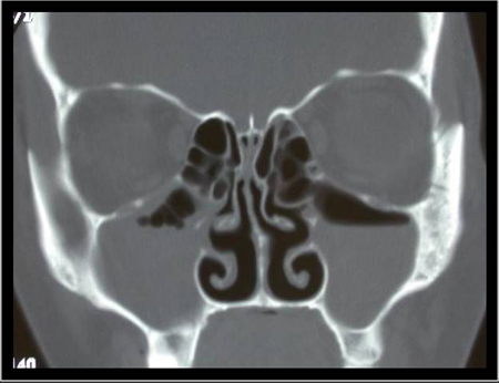
[Figure caption and citation for the preceding image starts]: Normal non-contrast computed tomography scan of the sinusesFrom the collection of Melissa Pynnonen, MD [Citation ends].
Result
identifies extent of sinus disease, abnormal anatomical structures
MRI
Test
MRI with and without intravenous contrast may be useful if extrasinus complications are suspected.[1][24] Not recommended for evaluation of routine, uncomplicated acute rhinosinusitis.[1]
MRI orbits, face, and neck without and with IV contrast can confirm paranasal sinus inflammation and identify orbital and adjacent intracranial complications.[24]
Result
may show air-fluid level or mucosal thickening of the involved sinuses, or extension of disease into adjacent structures such as the orbit or brain
lateral neck x-ray
Test
Lateral neck x-rays can be helpful in children to evaluate the patient for adenoid hypertrophy in patients with nasal obstruction.[15] An alternative is flexible nasal endoscopy, which can confirm adenoiditis.
Result
may show adenoid hypertrophy in children
allergy testing
Test
Skin or blood allergy testing (specific IgE) may be considered to rule out allergic rhinitis, particularly if there is no response to antibiotic therapy or for recurrent episodes.[1]
Result
positive
Use of this content is subject to our disclaimer