Investigations
1st investigations to order
electrophoresis with immunofixation and measurement of serum immunoglobulins
Test
The preferred test. Repeated after 6 months and then annually. Immunofixation identifies and measures the type of M protein. Patients with higher levels of M protein (>15g/L [1.5 g/dL] for IgG, and >10 g/L [1.0 g/dL] for IgA and IgM) are at greater risk for progression to myeloma. Non-IgG MGUS confers a higher risk for myeloma progression than IgG MGUS.[25][Figure caption and citation for the preceding image starts]: Using electrophoresis and immunofixation, figure A shows normal results while figure B shows evidence of an IgA kappa MGUS with polyclonal IgG backgroundFrom the collection of Ola Landgren, MD, PhD, courtesy of Dr Jerry Katzmann, Mayo Clinic, Rochester, MN [Citation ends].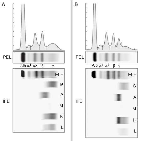
Result
positive for monoclonal (M) protein <30 g/L (3 g/dL)
FBC with differential
Test
Evaluates for anaemia, thrombocytopenia, and neutropenia.
Anaemia can be due to other causes, in which case further studies are required to rule out other underlying conditions.
By definition, the diagnosis of MGUS requires the absence of anaemia.
Result
normal
serum calcium
Test
In the presence of elevated calcium, other underlying conditions such as hyperparathyroidism should be ruled out.
By definition, the diagnosis of MGUS requires the absence of hypercalcaemia.
Result
normal
serum creatinine
Test
In the presence of elevated creatinine, other underlying conditions such as diabetes mellitus and hypertension should be ruled out.
By definition, the diagnosis of MGUS requires the absence of renal failure.
Result
normal
urinalysis and 24-hour urine collection with electrophoresis and immunofixation
Test
Dipstick of urine for protein is not sufficient. It primarily detects albumin, but it often does not detect immunoglobulin light chains.
Result
immunofixation identifies the isotype of monoclonal protein
serum free light chains assay
Test
Measures free circulating kappa and lambda light chains detectable in the blood.
Abnormal free light chain ratio indicates that the proportion of free circulating kappa and lambda light chains is abnormally high or low (equivalent to too much or too little kappa or lambda).
Abnormal ratio is an adverse risk factor for multiple myeloma progression.[29]
Result
normal: kappa-to-lambda ratio 0.26 to 1.65; abnormal: suggests work-up for multiple myeloma
metastatic bone survey (conventional x-ray or whole body low-dose CT scan)
Test
By definition, the diagnosis of MGUS requires the absence of lytic bone lesions related to plasma cell proliferative disorders.
A complete radiographic bone survey of the skeleton, including all long bones, should be performed using conventional x-ray or whole body low-dose CT scan if the monoclonal (M) protein level is high (>15g/L [1.5 g/dL] for IgG; >10 g/L [1.0 g/dL] for IgA and IgM); or if the patient has non-IgG MGUS; or if the free light chain ratio is abnormal.
Skeletal survey is also needed in patients who have unexplained anaemia or other cytopenias, renal failure, or hypercalcaemia.
Suspected metastases should prompt work-up for cancer, and lytic bone lesions a work-up for myeloma.
Result
negative
Investigations to consider
bone marrow aspiration and/or biopsy
Test
Should be performed if the monoclonal (M) protein is >15g/L (1.5 g/dL); or if the patient has a non-IgG MGUS; or if the free light chain ratio is abnormal.
Bone marrow should also be examined in patients who have unexplained anaemia or other cytopenias, renal failure, hypercalcaemia, or bone lesions on a metastatic bone survey. [Figure caption and citation for the preceding image starts]: Haematoxylin-eosin section of bone marrow biopsy showing mild increase in plasma cellsFrom the collection of Ola Landgren, MD, PhD [Citation ends].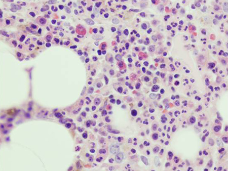 [Figure caption and citation for the preceding image starts]: CD138 immuno-histochemical staining highlighting small clusters of plasma cellsFrom the collection of Ola Landgren, MD, PhD [Citation ends].
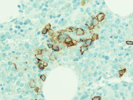
The probability of marrow having ≥10% plasma cells can be estimated using the iStopMM calculator, helping to predict the need for bone marrow sampling. iStopMM: predicting the need for bone marrow sampling in MGUS Opens in new window
Result
<10% abnormal plasma cells
bone mineral density scan
Test
Dual energy x-ray absorptiometry (DEXA) imaging may be used to assess bone mineral density, as patients with MGUS have an increased risk of osteoporosis.
Result
normal, or presence of osteoporosis
MRI scan
Test
MRI of the spine and pelvis may be used to evaluate bone pathology to help predict the likelihood of progression to multiple myeloma.
Result
normal; presence of bone lesions suggests a high risk of progression to multiple myeloma
PET scan
Test
PET imaging of the spine and pelvis may be used to evaluate bone pathology to help predict the likelihood of progression to multiple myeloma.
Result
normal; presence of bone lesions suggests a high risk of progression to multiple myeloma
Use of this content is subject to our disclaimer