Images and videos
Images


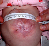


Omphalocele and gastroschisis
Note the membrane covering the abdominal contents in this omphalocele
From collection of J.J. Tepas III, MD, FACS, FAAP
See this image in context in the following section/s:
Omphalocele and gastroschisis
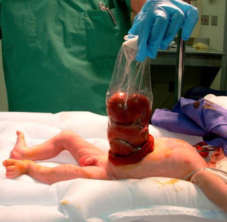
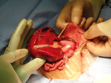
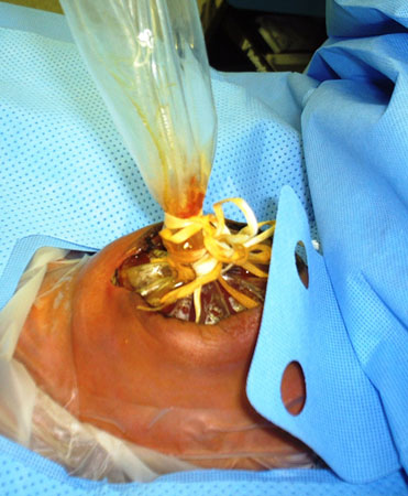
A staged repair of gastroschisis involves the placement of a silo to reduce contents into the abdomen
From collection of J.J. Tepas III, MD, FACS, FAAP
See this image in context in the following section/s:
Omphalocele and gastroschisis
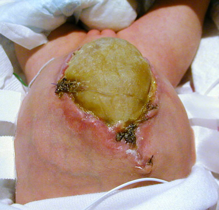
The abdominal wall defect in omphalocele is covered with a synthetic membrane
From collection of J.J. Tepas III, MD, FACS, FAAP
See this image in context in the following section/s:
Omphalocele and gastroschisis
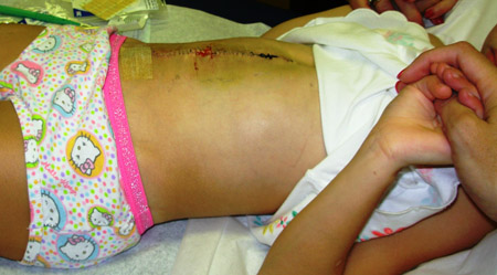
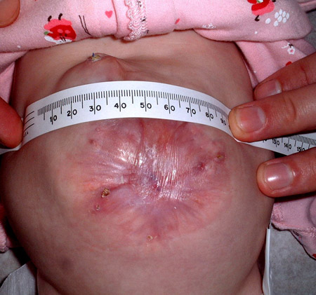
The ventral hernia is repaired in a 6-year-old girl born with omphalocele
From collection of J.J. Tepas III, MD, FACS, FAAP
See this image in context in the following section/s:
Omphalocele and gastroschisis
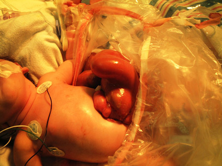
Immediately after delivery, an infant with gastroschisis is placed in a protective bowel bag
From collection of J.J. Tepas III, MD, FACS, FAAP
See this image in context in the following section/s:
Omphalocele and gastroschisis
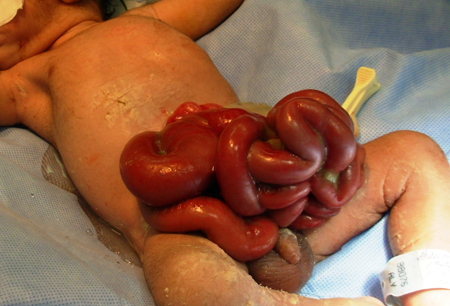
Extruded gut in abdominal wall defect
From collection of J.J. Tepas III, MD, FACS, FAAP
See this image in context in the following section/s:
Omphalocele and gastroschisis
This ruptured omphalocele is similar in appearance to gastroschisis
From collection of J.J. Tepas III, MD, FACS, FAAP
See this image in context in the following section/s:
Omphalocele and gastroschisis
A ventral hernia results from synthetic wall closure of the omphalocele
From collection of J.J. Tepas III, MD, FACS, FAAP
See this image in context in the following section/s:
Omphalocele and gastroschisis
Once intestinal contents are fully reduced into the abdomen, closure of the abdominal wall follows
From collection of J.J. Tepas III, MD, FACS, FAAP
See this image in context in the following section/s:
Use of this content is subject to our disclaimer