Investigations
1st investigations to order
echocardiography
Test
The single most important and useful test in the diagnosis and follow-up of patients with ventricular septal defects (VSDs).[2][11] Comprehensive transthoracic echocardiogram can also evaluate any concomitant heart defects.[22][23][24] 2D imaging and colour flow Doppler are usually diagnostic: the defect is clearly visualised on 2D views, and spectral-wave Doppler serves to confirm a high-velocity jet across the VSD demonstrating the left-to-right shunt.
The degree of shunting can be estimated by Doppler, and a pulmonary-systemic blood flow ratio (Qp:Qs) can be determined.
Also diagnoses associated conditions (e.g., tetralogy of Fallot) and complications (chamber enlargement, aortic regurgitation).
Continuous-wave Doppler of the tricuspid regurgitation jet can be used to assess the degree of pulmonary hypertension. [Figure caption and citation for the preceding image starts]: Echocardiographic image of a type 4 (muscular) ventricular septal defect (VSD) with colour flow Doppler showing left-to-right shuntFrom the collection of Dr Kul Aggarwal [Citation ends].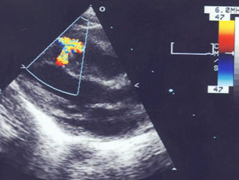 [Figure caption and citation for the preceding image starts]: Echocardiographic image with colour flow Doppler showing left-to-right shunting across a type 1 ventricular septal defect at the supracristal (or doubly committed juxta-arterial) levelFrom the collection of Dr Zuhdi Lababidi [Citation ends].
 [Figure caption and citation for the preceding image starts]: Echocardiographic image with colour flow Doppler showing left-to-right shunting across a type 3 ventricular septal defect at the inlet cushion levelFrom the collection of Dr Zuhdi Lababidi [Citation ends].
 [Figure caption and citation for the preceding image starts]: Echocardiographic image showing ventricular septal defect at the inlet cushion levelFrom the collection of Dr Zuhdi Lababidi [Citation ends].
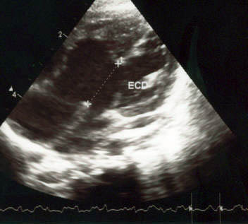 [Figure caption and citation for the preceding image starts]: Doppler image showing spectral recording of continuous-wave Doppler showing the left-to-right gradient across the ventricular septal defectFrom the collection of Dr Kul Aggarwal [Citation ends].
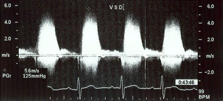
Result
VSD visualised and diagnosed on 2D views; left-to-right shunt confirmed by the presence of a high-velocity jet across the VSD on colour Doppler
chest x-ray
Test
In patients with small ventricular septal defects, the chest x-ray may be normal.
In patients with significant defects, there may be cardiomegaly and increased vascular markings.
In patients with Eisenmenger's syndrome, the chest x-ray may show a prominent pulmonary conus with peripheral pruning of the pulmonary vascular markings.[5]
Result
ranges from normal to cardiomegaly and increased pulmonary vascular markings to peripheral pruning of vascular markings
ECG
Test
Not diagnostic of a ventricular septal defect, but provides supportive evidence of chamber enlargement if present.
The ECG may be normal in small, non-restrictive defects.
In moderate-sized defects, there may be evidence of left ventricular and also sometimes left atrial enlargement.
In cases of larger shunts, there may be evidence of bi-ventricular hypertrophy.
In a few patients, there may be predominant right ventricular hypertrophy.
Result
various findings depending upon size of defect and the presence of pulmonary hypertension
Investigations to consider
cardiac MRI
Test
MRI and CT scan are complementary imaging modalities especially when other tests such as echocardiography are not optimal. MRI may be very useful in cases with complex defects or where echocardiography leaves some doubt.[21]
Result
visualisation of the defect; evaluation of the degree of shunting
cardiac CT scan
Test
MRI and CT scan are complementary imaging modalities especially when other tests such as echocardiography are not optimal.[21] Can be particularly useful in patients in whom MRI is contraindicated or if MRI shows artifacts from pacemaker leads, for example. Also has the advantage of allowing assessment of coronary arteries.
Result
may show location of ventricular septal defect, as well as chamber sizes and function
cardiac catheterisation
Test
Reserved for cases where a calculation of pressures and resistances is required, or non-invasive tests do not sufficiently define cardiac anatomy.[2]
Confirms the location of the shunt and the degree of pulmonary hypertension. Also serves to exclude significant concomitant coronary artery disease in older patients.
Measurement of pulmonary vascular resistance is used to assess suitability for surgical correction. [Figure caption and citation for the preceding image starts]: X-ray image showing a type 4 (muscular) ventricular septal defect on left ventriculographyFrom the collection of Dr Zuhdi Lababidi [Citation ends].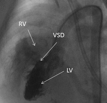 [Figure caption and citation for the preceding image starts]: X-ray image showing a type 2 (membranous) ventricular septal defect on left ventriculographyFrom the collection of Dr Zuhdi Lababidi [Citation ends].
Result
measurement of pulmonary artery pressures; demonstration of the location and degree of shunting
Use of this content is subject to our disclaimer