Approach
The diagnosis of an acute exacerbation is based on the history and clinical findings. A rapid triage assessment consisting of respiratory rate, heart rate, oxygen saturation, and temperature is made at the time of presentation to guide treatment. The examination should include chest auscultation, peak expiratory flow (PEF), and/or forced expiratory volume in the first second of expiration (FEV₁), but these should not unduly delay initial treatment. Exacerbations are then classified into mild, moderate, severe, or life-threatening.[7][57]
Please refer to the Diagnostic criteria for details of how to assess a child presenting with an acute exacerbation by age and severity.
This topic covers the treatment of children up to 11 years of age. Children 12 years and older are treated the same as adults (see Acute asthma exacerbation in adults).
History and assessment of risk factors
Assess the onset and duration of symptoms and any response to treatment, but be aware that children and parents often perceive severity inaccurately.[58] Most exacerbations occur in children known to have asthma. Children without a diagnosis of asthma may have a personal history of atopy or a first-degree relative with atopy (e.g., eczema, allergic rhinitis, and food allergies).
Key features of an acute exacerbation of asthma include the following:
A sudden or gradual increase in wheezing, coughing, chest tightness, and respiratory distress with lung hyper-inflation due to air trapping. Wheeze is a poor predictor of severity (e.g., silent chest in severe airflow obstruction) and cough is rarely the sole manifestation and should not be used as a marker of severity.
Exercise limitation and sleep disturbance may be reported.
Exposure to triggers such as viral infection, inhaled allergens, environmental irritants (e.g., tobacco smoke), emotions (e.g., anger, anxiety), the act of laughter, or exercise.
A previous history of asthma or atopy. Establish the patient's usual medication regimen, adherence to regular medications, and any changes made in response to the exacerbation (e.g., based on their self-management plan).
Risk factors for asthma-related death are also established. Factors include:[7]
A history of near-fatal asthma requiring intubation and mechanical ventilation
Hospitalisation or emergency care visit for asthma in the past year
Currently using or having recently stopped oral corticosteroids (a marker of event severity)
Not currently using inhaled corticosteroids (ICS)
Overuse of short-acting beta-2 agonists (SABA), especially use of more than one inhaler of salbutamol (or equivalent) monthly
Poor adherence with ICS containing medications and/or poor adherence to (or lack of) a written asthma action plan
A history of psychiatric disease or psychosocial problems
Suspected or confirmed anaphylaxis
Food allergy in a patient with asthma
Comorbidities (e.g., pneumonia, diabetes, and arrhythmias).
Physical examination
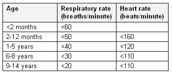
Perform a rapid triage assessment of respiratory rate, heart rate, oxygen saturation, and temperature:
Tachypnoea, commonly accompanied by tachycardia, is a characteristic sign of an acute exacerbation. Bradycardia is a pre-terminal sign.
Oxygen saturation may reveal the need for oxygen therapy. Cyanosis is a pre-terminal sign, indicative of severe hypoxaemia and respiratory failure.
Exhaustion and decreased consciousness are signs of impending respiratory arrest.
Pyrexia suggests infection.
[Figure caption and citation for the preceding image starts]: Normal ranges for respiratory rate and heart rate when awake at restCreated by the BMJ Evidence Centre with information from authors A. Chang and P. Robinson [Citation ends].
Look for evidence of respiratory distress:
The ability to speak is usually directly related to the degree of breathlessness but needs to be evaluated in relation to developmental age. In infants, the ability to feed reflects the degree of respiratory distress.
Intercostal, sub-costal, or sternal retraction may be present, reflecting the large negative pressures generated to increase air flow. Nasal flaring and tracheal tug may also be present.
Accessory muscles (e.g., sternocleidomastoid, parasternal, and scalene muscles) are recruited with more severe respiratory distress. In young infants, accessory muscle recruitment may manifest as head bobbing.
Auscultation of the lung fields typically reveals high-pitched, widespread, polyphonic wheeze, with a prolonged expiratory phase (i.e., usually an inspiratory-to-expiratory ratio of 1:2 or greater). The intensity of wheeze is a poor predictor of severity. Although wheeze may become biphasic with increasing airway obstruction, the absence of wheeze (silent chest) and the presence of pulsus paradoxus indicate a severe exacerbation.
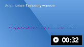
Auscultation sounds: Expiratory wheeze
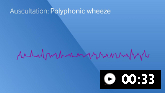
Auscultation sounds: Polyphonic wheeze
Pulsus paradoxus may be present (a fall >10 mmHg in systolic blood pressure with inspiration), but it is a poor indicator of exacerbation severity and is challenging to elicit in practice.
Response to treatment with a short-acting beta-2 agonist (SABA)
Reversible bronchoconstriction is a hallmark feature of asthma, and the initial response to bronchodilator therapy can be a useful guide to the accuracy of asthma as the diagnosis when objective confirmation is not possible.[59] A lack of response to SABA can indicate a severe asthma exacerbation, but should also prompt consideration of other diagnoses (e.g., recurrent viral wheeze), especially if seen in the context of stable, well-controlled asthma or as a first presentation of asthma-like symptoms.[60]
Initial investigations depending on severity
Oxygen saturation measured by pulse oximetry (SpO₂)
SpO₂ should be measured immediately.[7]
Significant hypoxaemia with an SpO₂ <90% is infrequent and, if present, represents severe airflow limitation and a severe or life-threatening exacerbation.
Pulse oximetry may overestimate oxygen saturation in people with dark skin colour.[7]
Reproducible spirometry and PEF
Rarely used during the initial assessment or monitoring of acute asthma exacerbations in children because they are difficult to assess, especially in young children, and stay normal in many attacks.
They may be useful in identifying alternative diagnoses in the differential.
If attempted, measurement should be performed with the patient seated and the best value of three attempts taken.
The degree of reduction in PEF, expressed as a percentage of the predicted value, reflects exacerbation severity. For children aged 6-11 years, a PEF >50% best or predicted indicates a mild or moderate exacerbation and a PEF ≤50% best or predicted indicates a severe or life-threatening exacerbation.[7]
Blood gases
Arterial blood gases (ABGs) are rarely measured in children and are reserved for life-threatening exacerbations.[7]
Free-flowing venous blood gases, taken at the time of intravenous cannulation, provide an approximation of partial pressure of carbon dioxide (PaCO₂).[61]
The typical finding is of low PaCO₂ due to pronounced tachypnoea initially (i.e., increased minute ventilation), but PaCO₂ can also be elevated in children with impending respiratory arrest.
Impending exhaustion and respiratory failure is associated with CO₂ retention
Other investigations
Chest x-ray is not indicated routinely. Its main use, to exclude other diagnoses, is indicated in patients presenting with their first episode of asthma, particularly if clinical features are atypical, and in patients with severe exacerbations who have had an atypical response to initial treatment. It is also indicated to assess focal signs on examination suggestive of pneumonia or pneumothorax.[7]
The chest x-ray is often normal, but findings may include hyper-inflation (in uncomplicated asthma), pneumothorax, atelectasis, pneumonia, or lobar collapse with consolidation.
Fractional exhaled nitric oxide (FeNO) is not currently recommended for assessing exacerbation severity or guiding acute asthma treatment.[7][55]
Where the equipment is available, NICE recommends FeNO as part of the initial treatment and assessment of acutely unwell children, but this recommendation is not consistent across current international guidelines.[62]
Use of this content is subject to our disclaimer