Images and videos
Images

Assessment of ataxia
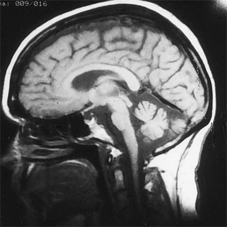
MRI of brain showing early cerebellar and brain stem atrophy in SCA 1
From the collection of Dr S. H. Subramony; used with permission
See this image in context in the following section/s:

Assessment of ataxia
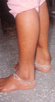
Tendon xanthoma seen in cerebrotendinous xanthomatosis
From the collection of Dr S. H. Subramony, with thanks to Dr Uday Muthane; used with permission
See this image in context in the following section/s:
Assessment of ataxia
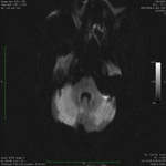
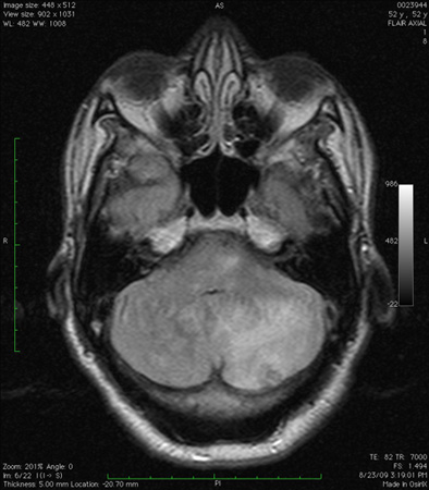
Cerebellar infarct as seen on fluid-attenuated inversion recovery sequence magnetic resonance image: note secondary oedema and effacement of the fourth ventricle
From the collection of Dr S. H. Subramony; used with permission
See this image in context in the following section/s:
Assessment of ataxia
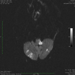
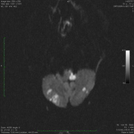
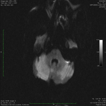
Infarct in the dorsolateral medulla together with scattered infarcts in the cerebellum, as seen on diffusion-weighted imaging sequence magnetic resonance image
From the collection of Dr S. H. Subramony; used with permission
See this image in context in the following section/s:
Assessment of ataxia
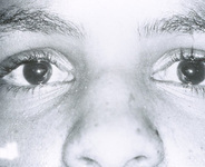
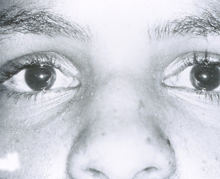
Early conjunctival telangiectasia in a patient with ataxia telangiectasia
From the collection of Dr S. H. Subramony; used with permission
See this image in context in the following section/s:
Assessment of ataxia
Acute bilateral cerebellar infarct, as seen on diffusion-weighted imaging sequence magnetic resonance image
From the collection of Dr S. H. Subramony; used with permission
See this image in context in the following section/s:
Assessment of ataxia
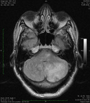
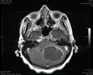
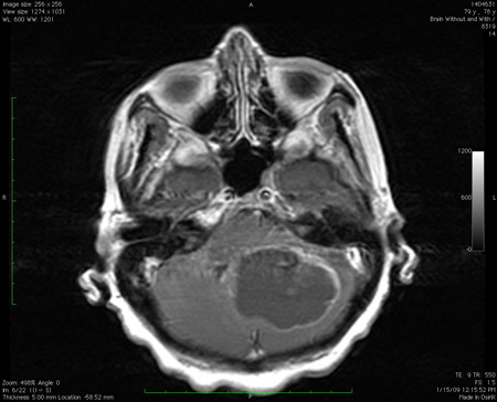
Large mass lesion in the cerebellum with pressure effects, as seen on MRI
From the collection of Dr S. H. Subramony; used with permission
See this image in context in the following section/s:
Assessment of ataxia
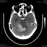
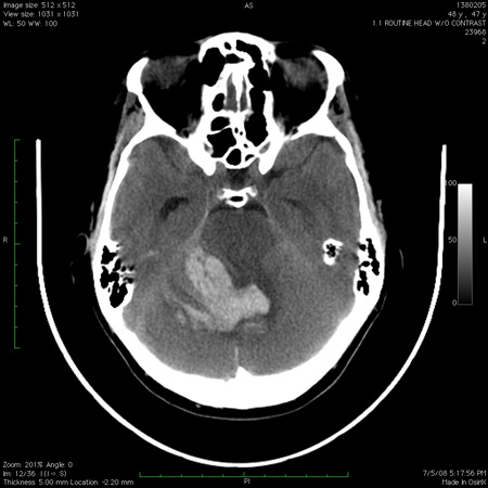
Computed tomography scan of the brain showing a haemorrhage in the cerebellum with extension into the fourth ventricle
From the collection of Dr S. H. Subramony; used with permission
See this image in context in the following section/s:

Assessment of ataxia
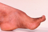
Pes cavus deformity in Friedreich's ataxia
From the collection of Dr S. H. Subramony; used with permission
See this image in context in the following section/s:
Use of this content is subject to our disclaimer