Images and videos
Images

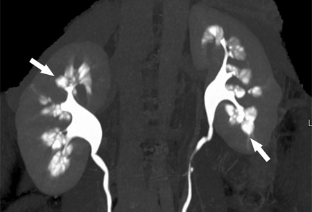
Medullary sponge kidney
Computed tomography (CT) urogram showing papillary blush with calculi within dilated collecting tubules (arrows)
From Maw AM, et al. Am J Kidney Dis. 2007 Jul;50(1):146-50, with permission
See this image in context in the following section/s:
Medullary sponge kidney
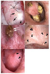
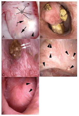
Endoscopic images of renal papilla from medullary sponge kidney (MSK) stone formers. The affected papillae are characterised by a rounding of contours and enlargement, which creates a billowy appearance (a and b). As part of the general rounding of contours, the papillary tips are blunted. These papillae also show sites of white (panel a, arrowhead) and yellow (a, arrows) plaque, and dilated opening of ducts of Bellini with (c, double arrow) and without deposits (a, asterisk). An occasional calyceal stone is noted (c). The unaffected papillae possess a normal morphology as seen in the compound papillum in (d) and single papillum in (e) (case 5); note numerous sites of white plaque (arrowheads) and an attached stone (double arrowhead). The unique morphology of affected papillae of MSK patients can be characteristic of all papillae (diffuse pattern) of a kidney or only some of the papillae (segmental pattern)
From Evan AP, et al. Anat Rec. 2015 May;298(5):865-77, with permission
See this image in context in the following section/s:
Use of this content is subject to our disclaimer