Images and videos
Images


Takayasu's arteritis
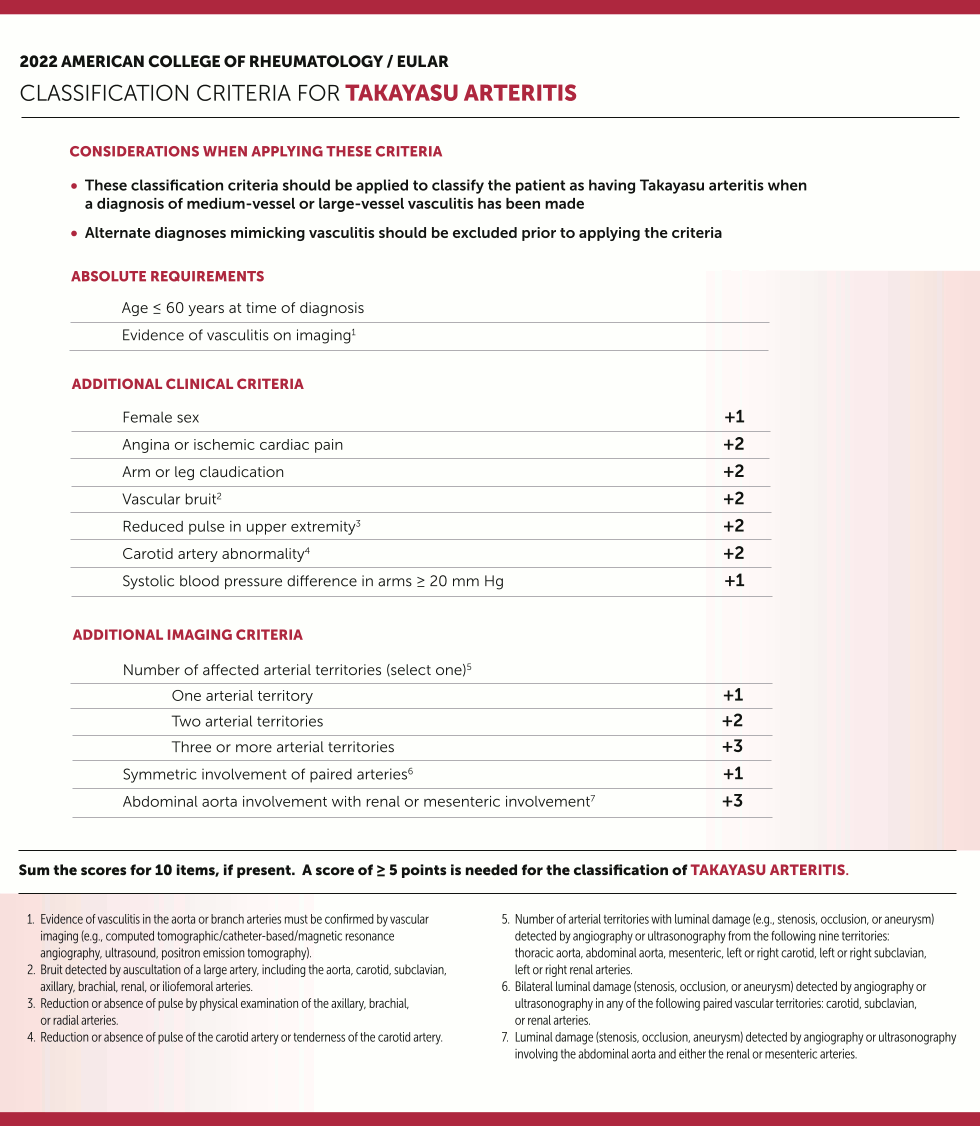
The final 2022 American College of Rheumatology/EULAR Classification Criteria for Takayasu arteritis
Grayson et al. Arthritis Rheumatol 2022;74(12):1872-80; used with permission
See this image in context in the following section/s:
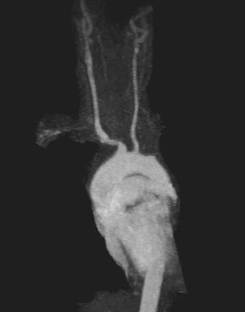
Takayasu's arteritis
Magnetic resonance angiogram of the aortic arch and major vessels showing occlusion of bilateral subclavian arteries; left common carotid artery has small diameter; proximal vertebral arteries are not identified
From the collection of Kenneth J. Warrington, MD
See this image in context in the following section/s:

Takayasu's arteritis
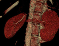
Computed tomography angiogram with 3D reconstruction showing bilateral renal artery stenosis
From the collection of Kenneth J. Warrington, MD
See this image in context in the following section/s:

Takayasu's arteritis
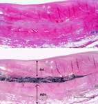
Photomicrograph of the aorta from a patient with Takayasu's arteritis demonstrates marked thickening of the intimal layer and inflammatory infiltrates in the media and laminar necrosis
Used with permission from the collection of Dylan Miller, MD, Mayo Clinic
See this image in context in the following section/s:

Takayasu's arteritis
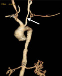
Computed tomography angiogram, with 3D reconstruction of the aortic arch and major vessels, showing proximal occlusion of the left subclavian artery and patent left vertebral artery distal to the occlusion (left vertebral steal syndrome)
From the collection of Kenneth J. Warrington, MD
See this image in context in the following section/s:
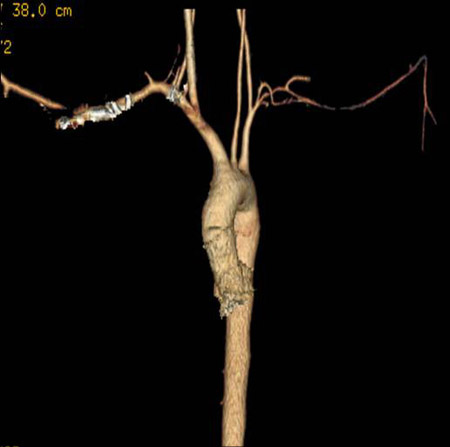
Takayasu's arteritis
Computed tomography angiogram, with 3D reconstruction of the aortic arch and major vessels, showing narrowing of the left common carotid artery and left subclavian artery
From the collection of Kenneth J. Warrington, MD
See this image in context in the following section/s:
Use of this content is subject to our disclaimer