Images and videos
Images


Hospital-acquired pneumonia (non COVID-19)
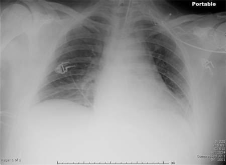
Portable chest x-ray of a patient with HAP. Note obscured left hemidiaphragm due to a left lower lobe opacity and an obscured heart border due to a left upper lobe or lingular opacity
Consent obtained at University of Louisville, KY
See this image in context in the following section/s:

Hospital-acquired pneumonia (non COVID-19)
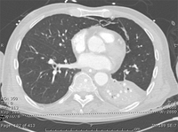
CT scan of a patient with a large, dense left lower lobe infiltrate
Consent obtained at University of Louisville, KY
See this image in context in the following section/s:
Hospital-acquired pneumonia (non COVID-19)
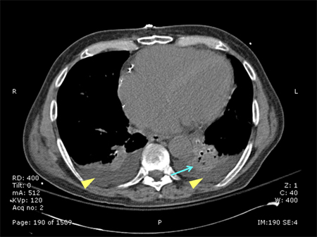
CT scan of a patient with a left lower lobe alveolar infiltrate (blue arrow), bilateral pleural effusions (yellow arrowheads), and right basilar atelectasis; notice the line separating the two shades of gray representing the infiltrate and the fluid
Consent obtained at University of Louisville, KY
See this image in context in the following section/s:

Hospital-acquired pneumonia (non COVID-19)
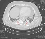
CT scan showing bibasilar opacities of patient with HAP
Consent obtained at University of Louisville, KY
See this image in context in the following section/s:
Videos
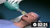
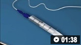 Supraglottic airway devices: animated demonstration
How to size and insert a laryngeal mask airway.
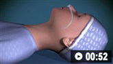 Nasopharyngeal airway: animated demonstration
How to select the correct size naspharyngeal airway and insert the airway device safely.
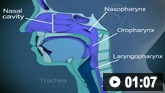 Oropharyngeal airway: animated demonstration
How to size and insert an oropharygeal airway.
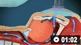 Bag-valve-mask ventilation: animated demonstration
How to use bag-valve-mask apparatus to deliver ventilatory support to adults. Video demonstrates the two-person technique.
 Tracheal intubation: animated demonstration
How to insert a tracheal tube in an adult using a laryngoscope.
Use of this content is subject to our disclaimer