Aetiology
Burkholderia mallei (glanders) and Burkholderia pseudomallei (melioidosis) are gram-negative, oxidase-positve bacilli that belong to the genus Burkholderia. They grow relatively easily on routine culture media, although B mallei is less robust than B pseudomallei. Selective media may be needed to isolate the organisms from sites with normal flora. They are sometimes discarded as contaminants by the unwary, although a characteristic sensitivity pattern (resistance to aminoglycosides and polymyxins but susceptibility to amoxicillin/clavulanate) is found in most B pseudomallei. B mallei is usually gentamicin susceptible. There have been reports of B pseudomallei that are susceptible to gentamicin in one specific geographical region, but this is unusual elsewhere.[36]
It is thought that melioidosis is usually acquired by inoculation of environmental organisms (e.g., through skin wounds acquired whilst working in rice paddies), but there is epidemiological evidence that it may also be acquired by inhalation, especially during heavy rainfall and severe weather events.[37][38] Ingestion of contaminated water may also be a previously under-recognised source of infection, especially in Southeast Asia.[39] In practice, it is usually very difficult to pinpoint precisely when, where, and how an individual became infected, particularly as people are being constantly exposed to the organism in the environment, and because incubation periods may be quite variable, a feature which has given rise to the nickname 'Vietnam time bomb'.[40]
Transmission of glanders to humans is through contact with tissues or body fluids of infected animals, or with cultures of B mallei in the laboratory. The bacteria enter the body through cuts or abrasions in the skin and through mucosal surfaces such as the eyes and nose. It may also be inhaled in infected aerosols or dust contaminated by infected animals.[35] Cases of human-to-human transmission are very rare.[41]
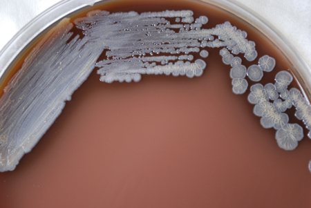
[Figure caption and citation for the preceding image starts]: Colonial morphology displayed by gram-negative Burkholderia pseudomallei bacteria (melioidosis), grown on a medium of chocolate agar, for a 72-hour time period, at a temperature of 37°CCDC Public Health Image Library (PHIL) [Citation ends]. [Figure caption and citation for the preceding image starts]: Colonial morphology displayed by gram-negative Burkholderia mallei bacteria (glanders), grown on a medium of chocolate agar, for a 72-hour time period, at a temperature of 37°CCDC Public Health Image Library (PHIL) [Citation ends].
Pathophysiology
The spectrum of clinical presentations reflects a range of possible outcomes of infection that depend on a balance between the size and route of initial inoculum and the effectiveness of the host response. Initial bacterial multiplication at the site of entry may lead to a local lesion such as skin sore with cellulitis, abscess formation, or pneumonia. In some patients this may resolve, but in others the organism may spread via the bloodstream or lymphatic system, leading to secondary lesions in virtually any site, but particularly in lymph nodes, liver, spleen, lung, and (in men) prostate.
The progression of disease may be very variable, with some patients developing acute, fulminating infection and others developing chronic, indolent, granulomatous lesions, which may occasionally be asymptomatic.[42] A number of virulence determinants have been described in Burkholderia mallei and Burkholderia pseudomallei, including an anti-phagocytic capsule, various secreted and cell-associated toxins, and a range of factors that enable organisms to survive intracellularly.[43]
Classification
Clinical classification of melioidosis[6]
1. Disseminated bacteraemic melioidosis
2. Non-disseminated bacteraemic melioidosis
3. Localised melioidosis
4. Transient bacteraemic melioidosis
5. Probable melioidosis
6. Subclinical melioidosis.
Melioidosis classification according to the presence/absence of septic shock and most prominent clinical focus of infection[7]
Septic shock:
Pneumonia
No evident focus (but may have internal organ abscesses on imaging)
Genitourinary
Osteomyelitis/septic arthritis
Soft tissue abscess.
Non-septic shock:
Pneumonia
Skin infection
Genitourinary
No evident focus
Soft tissue abscess(es)
Osteomyelitis/septic arthritis
Neurological.
Acute and chronic melioidosis[8]
Acute melioidosis (85%): incubation period 1 to 21 days (mean 9 days)
Chronic melioidosis (11%): defined as illness with symptoms for longer than 2 months' duration on presentation
Activation from latent focus from prior asymptomatic infection (4%).
Use of this content is subject to our disclaimer