Rib fractures are an indicator of severe trauma. Assessment of a patient suspected to have a rib fracture should therefore include identification of the fracture itself and any concurrent injuries. The proportion of patients with concomitant injuries can be as high as 90%.[31]Ziegler DW, Agarwal NN. The morbidity and mortality of rib fractures. J Trauma. 1994 Dec;37(6):975-9.
http://www.ncbi.nlm.nih.gov/pubmed/7996614?tool=bestpractice.com
The presence of multiple rib fractures correlates with an increased incidence of solid organ injury (about 35%).[26]Davoodabadi A, Mosavibioki N, Mashayekhil M, et al. Correlation of rib fracture patterns with abdominal solid organ injury: a retrospective observational cohort study. Chin J Traumatol. 2022 Jan;25(1):45-8.
https://www.ncbi.nlm.nih.gov/pmc/articles/PMC8787232
http://www.ncbi.nlm.nih.gov/pubmed/34303569?tool=bestpractice.com
Fracture of a first or second rib should alert the physician to the possibility of vascular or neurologic injury as greater force is required to fracture these two ribs, which are intimately associated with the subclavian artery and the brachial plexus.[23]Gupta A, Jamshidi M, Rubin JR. Traumatic first rib fracture: is angiography necessary? A review of 730 cases. Cardiovasc Surg. 1997 Feb;5(1):48-53.
http://www.ncbi.nlm.nih.gov/pubmed/9158123?tool=bestpractice.com
[24]Fermanis GG, Deane SA, Fitzgerald PM. The significance of first and second rib fractures. Aust N Z J Surg. 1985 Aug;55(4):383-6.
http://www.ncbi.nlm.nih.gov/pubmed/3870169?tool=bestpractice.com
[25]Poole GV. Fracture of the upper ribs and injury to the great vessels. Surg Gynecol Obstet. 1989 Sep;169(3):275-82.
http://www.ncbi.nlm.nih.gov/pubmed/2672391?tool=bestpractice.com
History and exam
Rib fractures are most commonly a result of motor vehicle collisions (MVCs), falls, blunt trauma, assaults, and industrial incidents.[6]Holcomb JB, McMullin NR, Kozar RA, et al. Morbidity from rib fractures increases after age 45. J Am Coll Surg. 2003 Apr;196(4):549-55.
http://www.ncbi.nlm.nih.gov/pubmed/12691929?tool=bestpractice.com
[8]Sirmali M, Türüt H, Topçu S, et al. A comprehensive analysis of traumatic rib fractures: morbidity, mortality and management. Eur J Cardiothorac Surg. 2003 Jul;24(1):133-8.
https://academic.oup.com/ejcts/article/24/1/133/375481
http://www.ncbi.nlm.nih.gov/pubmed/12853057?tool=bestpractice.com
[30]Kent R, Woods W, Bostrom O. Fatality risk and the presence of rib fractures. Annu Proc Assoc Adv Automot Med. 2008 Oct;52:73-82.
http://www.ncbi.nlm.nih.gov/pubmed/19026224?tool=bestpractice.com
Other risk factors include age >65 years, osteoporosis, and cardiopulmonary resuscitation.[4]Siris ES, Brenneman SK, Barrett-Connor E, et al. The effect of age and bone mineral density on the absolute, excess, and relative risk of fracture in postmenopausal women aged 50-99: results from the National Osteoporosis Risk Assessment (NORA). Osteoporos Int. 2006;17(4):565-74.
http://www.ncbi.nlm.nih.gov/pubmed/16392027?tool=bestpractice.com
[7]Marini CP, Petrone P, Soto-Sánchez A, et al. Predictors of mortality in patients with rib fractures. Eur J Trauma Emerg Surg. 2021 Oct;47(5):1527-34.
https://link.springer.com/article/10.1007/s00068-019-01183-5
http://www.ncbi.nlm.nih.gov/pubmed/31324938?tool=bestpractice.com
[13]Hoke RS, Chamberlain D. Skeletal chest injuries secondary to cardiopulmonary resuscitation. Resuscitation. 2004 Dec;63(3):327-38.
http://www.ncbi.nlm.nih.gov/pubmed/15582769?tool=bestpractice.com
[28]Kim EY, Yang HJ, Sung YM, et al. Multidetector CT findings of skeletal chest injuries secondary to cardiopulmonary resuscitation. Resuscitation. 2011 Jun 12;82(10):1285-8.
http://www.ncbi.nlm.nih.gov/pubmed/21705131?tool=bestpractice.com
The patient or the emergency medical service personnel can provide important information regarding the likely etiology, such as blunt trauma in a car accident. Significant intrusion of the steering column can apply dramatic force to the thoracic cage.
Pain and dyspnea are common. Chest wall pain reduces ventilation by impaired inspiratory effort. Impaired oxygenation can also be indicative of underlying pneumothorax, hemothorax, or pulmonary contusion. Paradoxical chest wall motion with inspiration or expiration is a sign of a flail chest. A flail chest results when multiple ipsilateral ribs are fractured in two or more places resulting in an unstable segment of the chest wall. Flail chest is often accompanied by other injuries and carries an increased risk of life-threatening respiratory failure, pneumothorax, pulmonary contusion, and hemothorax, with an overall mortality of at least 5%.[32]Athanassiadi K, Gerazounis M, Theakos N. Management of 150 flail chest injuries: analysis of risk factors affecting outcome. Eur J Cardiothorac Surg. 2004 Aug;26(2):373-6.
http://ejcts.oxfordjournals.org/content/26/2/373.long
http://www.ncbi.nlm.nih.gov/pubmed/15296899?tool=bestpractice.com
Other factors to consider:
Stress fractures occur in 2% to 12% of rowers as a result of cyclic loading to the rib cage.[22]Smoljanović T, Bojanić I, Troha I, et al. Rib stress fractures in rowers: three case reports and review of literature [in Croatian]. Lijec Vjesn. 2007 Oct-Nov;129(10-11):327-32.
http://www.ncbi.nlm.nih.gov/pubmed/18257332?tool=bestpractice.com
It is also common in overhead repetitive athletic activities, such as baseball and golf.[21]Gerrie BJ, Harris JD, Lintner DM, et al. Lower thoracic rib stress fractures in baseball pitchers. Phys Sportsmed. 2015 Nov 26;44(1):93-6.
http://www.ncbi.nlm.nih.gov/pubmed/26559562?tool=bestpractice.com
Diagnosis is suggested by continued pain despite the absence of trauma.
Anyone with a known malignancy, particularly of lung, prostate, breast, liver, or gastrointestinal origin, could present with rib metastasis.
Primary bone tumors of the chest wall, including osteochondroma, enchondroma, plasmacytoma, chondrosarcoma, and osteosarcoma, are rare but can manifest as rib fractures. About 37% of these lesions are malignant.[16]Aydoğdu K, Findik G, Agackiran Y, et al. Primary tumors of the ribs; experience with 78 patients. Interact Cardiovasc Thorac Surg. 2009 Aug;9(2):251-4.
https://academic.oup.com/icvts/article/9/2/251/729805
http://www.ncbi.nlm.nih.gov/pubmed/19447801?tool=bestpractice.com
Multiple myeloma can present with rib fractures and even with a flail chest.[17]Muñoz-Bermúdez R, Abella E, Zuccarino F, et al. Successfully non-surgical management of flail chest as first manifestation of multiple myeloma: a case report. World J Crit Care Med. 2019 Sep 11;8(5):82-6.
https://www.ncbi.nlm.nih.gov/pmc/articles/PMC6753394
http://www.ncbi.nlm.nih.gov/pubmed/31559147?tool=bestpractice.com
Any rib fracture in a child or infant should be assumed to be the result of nonaccidental trauma until shown otherwise. Among infants younger than 12 months presenting with rib fractures, as many as 82% sustained these injuries non-accidentally (i.e., through physical abuse).[2]Bulloch B, Schubert CJ, Brophy PD, et al. Cause and clinical characteristics of rib fractures in infants. Pediatrics. 2000 Apr;105(4):E48.
http://pediatrics.aappublications.org/content/105/4/e48.full
http://www.ncbi.nlm.nih.gov/pubmed/10742369?tool=bestpractice.com
[3]Paine CW, Fakeye O, Christian CW, et al. Prevalence of Abuse Among Young Children With Rib Fractures: A Systematic Review. Pediatr Emerg Care. 2019 Feb;35(2):96-103.
http://www.ncbi.nlm.nih.gov/pubmed/27749806?tool=bestpractice.com
Of all skeletal injuries, rib fractures have the highest likelihood of being the result of physical abuse.[14]Kemp AM, Dunstan F, Harrison S, et al. Patterns of skeletal fractures in child abuse: systematic review. BMJ. 2008 Oct 2;337:a1518.
http://www.ncbi.nlm.nih.gov/pmc/articles/PMC2563260/?tool=pubmed
http://www.ncbi.nlm.nih.gov/pubmed/18832412?tool=bestpractice.com
Investigations
Chest radiography is the first-line imaging modality in any patient presenting with known trauma.[33]American College of Radiology. ACR appropriateness criteria: rib fractures. 2018 [internet publication].
https://acsearch.acr.org/docs/69450/Narrative
[34]American College of Surgeons; Trauma Quality Improvement Program. Best practices guidelines in imaging. Oct 2018 [internet publication].
https://www.facs.org/media/oxdjw5zj/imaging_guidelines.pdf
This not only helps detect the actual rib fracture, but pneumothorax, hemothorax, and aortic injury can also be rapidly assessed. However, conventional chest x-rays (CXR) can miss up to 50% of rib fractures.[35]Livingston DH, Shogan B, John P, et al. CT diagnosis of rib fractures and the prediction of acute respiratory failure. J Trauma. 2008 Apr;64(4):905-11.
http://www.ncbi.nlm.nih.gov/pubmed/18404055?tool=bestpractice.com
In practice, a single rib fracture might be diagnosed clinically based on symptoms and a clear history of thoracic trauma, even if no fracture is identified on CXR; further imaging is only warranted if it has the potential to change patient management. It is not usually necessary to perform dedicated rib radiography, in addition to chest radiography, for the diagnosis of rib fractures in adults after minor trauma.[33]American College of Radiology. ACR appropriateness criteria: rib fractures. 2018 [internet publication].
https://acsearch.acr.org/docs/69450/Narrative
[Figure caption and citation for the preceding image starts]: CXR showing right first rib fractureFrom the collection of Dr Paul Novakovich; used with permission [Citation ends]. [Figure caption and citation for the preceding image starts]: CXR showing multiple posterior left-sided rib fracturesFrom the collection of Dr Paul Novakovich; used with permission [Citation ends].
 [Figure caption and citation for the preceding image starts]: Anteroposterior CXR multiple left-sided rib fractures with chest tube in placeFrom the collection of Dr Paul Novakovich; used with permission [Citation ends].
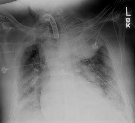
Computed tomography (CT) of the chest has greater sensitivity than CXR in detecting rib fractures as well as other injuries. A majority of patients who sustain major trauma and have evidence of chest injury on plain x-ray have an unsuspected injury identified on chest CT; up to one-third of these patients have their management significantly changed as a result of the chest CT.[36]Guerrero-López F, Vázquez-Mata G, Alcázar-Romero PP, et al. Evaluation of the utility of computed tomography in the initial assessment of the critical care patient with chest trauma. Crit Care Med. 2000 May;28(5):1370-5.
http://www.ncbi.nlm.nih.gov/pubmed/10834680?tool=bestpractice.com
CT imparts significant radiation exposure to the patient but should be considered if clinical features are suggestive of fracture and there is the potential for improved patient care if rib fracture(s) are detected. In minor trauma, however, the increased sensitivity of CT does not necessarily alter management or clinical outcomes of patients who do not have associated injuries.[33]American College of Radiology. ACR appropriateness criteria: rib fractures. 2018 [internet publication].
https://acsearch.acr.org/docs/69450/Narrative
[Figure caption and citation for the preceding image starts]: CT scan showing large left-sided pneumothoraxFrom the collection of Dr Paul Novakovich; used with permission [Citation ends].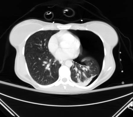 [Figure caption and citation for the preceding image starts]: CXR depicting the same pneumothorax as shown on CTFrom the collection of Dr Paul Novakovich; used with permission [Citation ends].
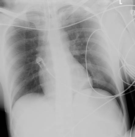 [Figure caption and citation for the preceding image starts]: CT scan showing bilateral posterior rib fracturesFrom the collection of Dr Paul Novakovich; used with permission [Citation ends].
 [Figure caption and citation for the preceding image starts]: CT showing right anterolateral rib fractureFrom the collection of Dr Paul Novakovich; used with permission [Citation ends].
 [Figure caption and citation for the preceding image starts]: CT showing left posterior segmental rib fractureFrom the collection of Dr Paul Novakovich; used with permission [Citation ends].
Ultrasound has been evaluated for its usefulness in diagnosing rib fractures. It is marginally superior to chest radiography, but may be associated with patient discomfort. Ultrasound is more typically used as an adjunct, in some centers, to rapidly assess for pneumothorax and pleural fluid; diagnostic accuracy is operator-dependent.[33]American College of Radiology. ACR appropriateness criteria: rib fractures. 2018 [internet publication].
https://acsearch.acr.org/docs/69450/Narrative
[37]Hurley ME, Keye GD, Hamilton S. Is ultrasound really helpful in the detection of rib fractures? Injury. 2004 Jun;35(6):562-6.
http://www.ncbi.nlm.nih.gov/pubmed/15135274?tool=bestpractice.com
[38]Stengel D, Leisterer J, Ferrada P, et al. Point-of-care ultrasonography for diagnosing thoracoabdominal injuries in patients with blunt trauma. Cochrane Database Syst Rev. 2018 Dec 12;12(12):CD012669.
https://www.ncbi.nlm.nih.gov/pmc/articles/PMC6517180
http://www.ncbi.nlm.nih.gov/pubmed/30548249?tool=bestpractice.com
Routine use of angiography is not indicated in the absence of discrepant pulses, mediastinal widening on CXR, brachial plexus injury, or an expanding hematoma.[23]Gupta A, Jamshidi M, Rubin JR. Traumatic first rib fracture: is angiography necessary? A review of 730 cases. Cardiovasc Surg. 1997 Feb;5(1):48-53.
http://www.ncbi.nlm.nih.gov/pubmed/9158123?tool=bestpractice.com
However, it may be used if there is a fracture of a first rib as traumatic injuries to the first rib are associated with a 3% risk of concomitant great-vessel injury.[23]Gupta A, Jamshidi M, Rubin JR. Traumatic first rib fracture: is angiography necessary? A review of 730 cases. Cardiovasc Surg. 1997 Feb;5(1):48-53.
http://www.ncbi.nlm.nih.gov/pubmed/9158123?tool=bestpractice.com
Typically, patients presenting after high-energy trauma should receive an immediate chest and pelvic radiograph to rule out life-threatening injury.[39]National Institute for Health and Care Excellence. Major trauma: assessment and initial management. Feb 2016 [internet publication].
https://www.nice.org.uk/guidance/ng39/chapter/Recommendations
If the patient is stable, a CT scan of the head, cervical spine, chest, abdomen, and pelvis may be performed in adults to rule out other injury.[39]National Institute for Health and Care Excellence. Major trauma: assessment and initial management. Feb 2016 [internet publication].
https://www.nice.org.uk/guidance/ng39/chapter/Recommendations
A skeletal survey and a consultation with child protective services should be considered in all children with suspected physical abuse. Oblique views of the ribs are recommended in all cases where abuse is suspected.[40]American College of Radiology. ACR appropriateness criteria: suspected physical abuse - child. 2016 [internet publication].
https://acsearch.acr.org/docs/69443/Narrative
Oblique views increase diagnostic accuracy for rib fractures, which are strong positive predictors of abuse, and may be the only skeletal manifestation.