Recommendations
Urgent
Assess the patient using the Airway, Breathing, Circulation, Disability, Exposure (ABCDE) approach.[32]
Recognise anaphylaxis by the sudden onset and rapid progression of symptoms: life-threatening Airway and/or Breathing and/or Circulation problems with or without skin and/or mucosal changes after exposure to a trigger (allergen).[1][32]
Give intramuscular adrenaline (epinephrine) in the anterolateral thigh and call for senior help.[32]
Most reactions develop over several minutes.[32]
Generalised urticaria, angio-oedema, and rhinitis without life-threatening airway, breathing, or circulation problems do not meet the criteria for anaphylaxis. But if in doubt, give intramuscular adrenaline and seek expert help.[32]
Airway:[32]
Throat and tongue swelling with difficulty breathing and swallowing; patient feels that their throat is ‘closing up’
Hoarse voice
Stridor.
Breathing:[32]
Increased respiratory rate
Wheeze
Hypoxia (oxygen saturation [SpO 2] <92%)
Confusion
Patient feels tired
Cyanosis (usually a late sign)
Respiratory arrest (may be the primary presentation).
Circulation:[32]
Signs of shock
Tachycardia
Low blood pressure with feeling of faintness/dizziness or collapse[32]
Adults and children aged 11 years or older: systolic blood pressure <90 mmHg or >30% decrease from baseline
Children aged 1 to 10 years: <70 mmHg + (2 x age in years)
Infants aged 1 to 12 months: <70 mmHg
Neonates aged <1 month: 50 to 60 mmHg
Decreased level of consciousness or loss of consciousness (due to decreased brain perfusion)
Chest pain with ECG changes (even in people with normal coronary arteries)[50]
Bradycardia and cardiac arrest.
Disability:
Confusion, agitation, and loss of consciousness associated with anaphylaxis can be caused by profound hypoxia, hypercapnia, or cerebral hypoperfusion[32]
May also be triggered by administration of adrenaline, but tends to resolve as the other allergic symptoms abate.
Exposure:[32]
While respecting the patient’s dignity, fully expose their body to identify skin reactions
These may be subtle or dramatic and are often the first sign of anaphylaxis[32]
Skin and/or mucosal changes (flushing, urticaria, angio-oedema) occur in 80% of patients with anaphylaxis.[7][32] However, absence of changes does not rule it out
Most patients with allergic skin changes do not progress to anaphylaxis[32]
Exposure to a trigger supports the diagnosis, but lack of obvious exposure does not exclude the diagnosis.[32] Common allergens include:
Food
Medications
Venoms
Latex
Exercise
Cold.
Patients are often anxious and may report a ‘sense of doom’.[32]
Consider serious differential diagnoses early.
Severe asthma may also present with wheezing, coughing, and shortness of breath. However, associated itching, urticaria, angio-oedema, abdominal pain, and hypotension is more suggestive of anaphylaxis.[51]
Sepsis can manifest with low diastolic blood pressure. However, associated skin changes are more likely to be petechial or purpuric in sepsis, compared with the erythema (patchy, or generalised, red rash) or urticaria (also called hives, nettle rash, wheals, or welts) associated with anaphylaxis.[32]
Seek senior help early if you are unsure of the diagnosis.
Key Recommendations
[Figure caption and citation for the preceding image starts]: Anaphylaxis diagnostic and management flowchart. See Management section for further information, including actions to take if there is no improvement in breathing or circulation problems after two doses of adrenaline.Created by the BMJ Knowledge Centre; adapted with permission from the Resuscitation Council (UK). Emergency treatment of anaphylaxis: guidelines for healthcare providers. May 2021 [Citation ends].
Assessment
Perform a quick general assessment to see whether the patient ‘looks unwell’.
Normal responses to a question or stimulation (if the patient appears unconscious) indicate a patent airway and brain perfusion.
Short sentences signal breathing problems.
Failure to respond suggests critical illness.[32]
Airway obstruction
Look for airway obstruction and treat as an emergency if present:[32]
Stridor
Paradoxical chest and abdominal movements and accessory muscles of respiration
No breath sounds, diminished air entry, or noisy air entry.
Call for expert help immediately, as airway swelling often requires early intubation, which may be technically challenging.[32]
Bronchospasm
Recognise acute severe bronchospasm.[32]
Respiratory distress is indicated by raised respiratory rate, sweating, central cyanosis, use of accessory respiratory muscles, subcostal and sternal recession in children, or abdominal breathing.[32]
Normal respiratory rates in breaths per minute:[32]
Adults and children >12 years: 12 to 20
Children
>5 to 12 years: 20 to 24
>2 to 5 years: 24 to 30
>1 to 2 years: 26 to 34
<1 year: 30 to 40.
Wheeze is commonly a feature of bronchospasm in anaphylaxis.[32]
Circulatory dysfunction
Identify shock.
Blue, pale, mottled, and cold hands and digits suggest circulatory collapse.[32]
These signs can be present in any pathological process that causes hypovolaemia, including sepsis.
Measure capillary refill time (normal is <2 seconds).
Veins may be under-filled or collapsed in hypovolaemia.[32]
Tachycardia is associated with circulatory collapse.[32]
Blood pressure may be normal or low.
Lower limit of systolic blood pressure (mmHg by age):[32]
Adults and children >10: 90
Children >1 to 10 years: 70 + (2 x age in years)
Children >1 to 12 months: 70
Newborn to 1 month: 50 to 60.
Reduced level of consciousness.[32]
Investigations
Measure blood mast cell tryptase concentrations in people aged 16 years and over as soon as possible after emergency treatment has started.[52][Evidence C]
Presentation is varied and the signs and symptoms are not specific for anaphylaxis. However, certain combinations make the diagnosis more likely.[32][53]
The diagnosis of anaphylaxis is clinical because management cannot wait for laboratory confirmation of allergy.
Consider serious differential diagnoses and seek senior help early if you are unsure of the diagnosis.
Severe asthma may also present with wheezing, coughing, and shortness of breath. However, associated itching, urticaria, angio-oedema, abdominal pain, and hypotension is more suggestive of anaphylaxis.[51]
Sepsis can manifest with low diastolic blood pressure. However, associated skin changes are more likely to be petechial or purpuric in sepsis, compared with the erythema (patchy, or generalised, red rash) or urticaria (also called hives, nettle rash, wheals, or welts) associated with anaphylaxis.[32]
Initial assessment
Perform a quick general assessment to see whether the patient ‘looks unwell’.
Normal responses to a question or stimulation (if the patient appears unconscious) indicate a patent airway and brain perfusion.
Short sentences signal breathing problems.
Failure to respond suggests critical illness.[32]
Base your diagnosis of anaphylaxis on the following three criteria:[1][32]
Sudden onset and rapid progression of symptoms, usually developing over several minutes to a maximum of 1 hour after exposure.[32] The patient is often anxious and can experience a sense of impending doom (angor animi).
Life-threatening Airway and/or Breathing and/or Circulation problems
Airway:
Throat and tongue swelling with difficulty breathing and swallowing; patient feels that their throat is ‘closing up
Hoarse voice
Stridor
Breathing:
Increased respiratory rate
Wheeze
Confusion caused by hypoxia (oxygen saturation [SpO 2] <92%)
Patient feels tired
Cyanosis (usually a late sign)
Respiratory arrest (may be the primary presentation).
Circulation:
Signs of shock
Tachycardia
Low blood pressure with feeling of faintness/dizziness or collapse[32]
Adults and children aged 11 years or older: systolic blood pressure <90 mmHg or >30% decrease from baseline
Children aged from 1 to 10 years: <70 mmHg + [2 x age in years]
Infants aged 1 to 12 months: <70 mmHg
Neonates aged <1 month: 50 to 60 mmHg
Decreased consciousness level or loss of consciousness (due to poor brain perfusion)
Chest pain with ECG changes (even in people with normal coronary arteries)
Bradycardia and cardiac arrest.
Skin and/or mucosal changes
[Figure caption and citation for the preceding image starts]: UrticariaCNRI/Science Photo Library; used with permission [Citation ends].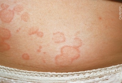
Practical tip
The lack of consistent clinical presentations makes anaphylaxis a diagnostic challenge.[32]
Be careful not to miss anaphylaxis in the absence of hypotension. This is an uncommon early presentation in children.[55][56]
Urticaria can be a feature of viral infections or febrile illnesses.
Life-threatening food allergy can trigger asthma without other features of anaphylaxis.[32][57]
Anaphylaxis can present as primary respiratory arrest.[32][54]
Exposure to a trigger supports the diagnosis.[32] However, a lack of obvious exposure does not exclude the diagnosis. See our History section below for common triggers.
Many patients are unaware of their food allergy and/or may be unaware that a particular food includes the allergen.
Further assessment
Having identified anaphylaxis using the three criteria outlined above, continue with the D and E elements of the ABCDE assessment.
Disability
Airway, breathing, and circulatory problems can affect neurological status leading to confusion, agitation, and loss of consciousness.[32]
May also be triggered by administration of adrenaline (epinephrine), but tends to resolve as the other allergic symptoms abate.
Gastrointestinal symptoms (abdominal pain, nausea, vomiting, incontinence) may also occur.[32] May occur together or singly and suggest allergen ingestion.
Exposure
While respecting the patient’s dignity, fully expose their body to identify skin reactions.
Skin/mucosal changes are often the first presenting feature and occur in over 80% of anaphylactic reactions, but their absence does not exclude the diagnosis of anaphylaxis.[7]
Erythema (patchy or generalised red rash) and urticaria (hives, wheals, or welts of different shapes/sizes) anywhere on the body (usually very itchy) are associated with anaphylaxis.[32]
Angio-oedema involves swelling of deeper tissues including eyelids, lips, mouth, and throat.
Most patients with allergic skin changes do not progress to anaphylaxis.[32]
Patients are often anxious and may report a ‘sense of doom’.[32]
Practical tip
Generalised urticaria, angio-oedema, and rhinitis do not meet criteria for anaphylaxis because life-threatening features – an airway problem, respiratory difficulty, and/or hypotension (circulatory compromise) – are absent. But if in doubt, give intramuscular adrenaline and seek expert help.[32]
Seek the following information from the patient, or from their carer if they are unable to communicate because of the severity of their symptoms.
Question | What the answer adds | Specific actions to take |
Do you have an allergy to known triggers and have you been exposed to a trigger (see table below)? | Supports the diagnosis. Nut allergies are thought to be associated with increased risk for a severe reaction.[32][58] | If signs of ABC difficulties develop, give adrenaline (epinephrine). Otherwise give antihistamine and observe carefully. |
Any previous episodes of anaphylaxis? | Bear in mind that the severity of the allergic response during each episode is often unpredictable.[43] | Have a lower threshold for giving adrenaline if you know the patient has a proven history of anaphylaxis to a specific trigger. |
Has adrenaline been administered on this or previous occasions? How does the patient tend to react to a particular trigger? | Provides information on the severity of previous allergic reactions. Some patients have severe cutaneous reactions, but don’t need adrenaline. However, the development of symptoms, and therefore the need for adrenaline, is unpredictable. | Have a lower threshold for giving adrenaline if you know the patient has a proven history of anaphylaxis to a specific trigger.[59] |
Where and when did the incident take place? | The patient’s location when symptoms started may provide clues: for example, restaurant (food allergy), garden (stings). An acute history with rapid, life-threatening symptom progression points to anaphylaxis. | Remove any food that may still be in the patient’s mouth, but do not induce vomiting.[32] Remove stings; removal is more important than the method of removal.[32] |
Are there other signs of atopy including asthma, eczema, and hayfever? | Supports, but does not define, the diagnosis.[2][38][39][40] May indicate a differential diagnosis. | If the patient has a history of asthma, have a lower threshold for giving adrenaline if there are other life-threatening signs.[32] Note that poorly controlled asthma increases the risk of death from anaphylaxis.[60][61] |
Are there any other concomitant diseases and medication? | Be particularly cautious; co-existing cardiovascular disease increases the risk of fatal anaphylaxis.[62] Beta-blockers may make treating anaphylaxis more difficult because they block the effect of adrenaline. Seek advice from a cardiologist, but do not delay giving adrenaline in a life-threatening anaphylaxis when adrenaline is required.[63] Mast cell disorders increase the severity and fatality.[53][64] Seek advice from a senior clinician urgently. | Seek advice from a senior clinician. |
Co-factors may be present in up to 20% of young people. They may increase the risk of an allergic reaction occurring or may make a reaction more severe.[59][68] | Be particularly cautious and observe the patient carefully for developing life-threatening symptoms. |
Common anaphylaxis triggers |
Food (in alphabetical order):[32][69]
Note that prevalence is greatest in children and decreases with age.[41] Anaphylaxis can also be caused by handling or inhalation of the food aerosol. |
|
Venoms (including bites and stings)[32] |
Latex:[32] |
Exercise:[32]
|
Hot or cold exposure: |
Idiopathic (unknown cause)[32] |
Carry out your initial assessment (see Clinical presentation above) and examination simultaneously as anaphylaxis is an acute life-threatening condition. In particular, look for life-threatening features including airway obstruction (A), bronchospasm (B), and circulatory dysfunction (C).
Airway obstruction
Look for airway obstruction and treat as an emergency if present:[32]
Stridor
Paradoxical chest and abdominal movements and accessory muscles of respiration
No breath sounds, diminished air entry, or noisy air entry.
Call for expert help from an anaesthetist immediately, as airway swelling often requires early intubation, which may be technically challenging.[32]
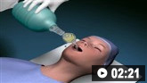
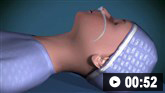
How to insert a tracheal tube in an adult using a laryngoscope.
Bronchospasm
Recognise acute severe bronchospasm.[32]
Respiratory distress is indicated by raised respiratory rate, sweating, central cyanosis, use of accessory respiratory muscles, subcostal and sternal recession in children, and abdominal breathing.[32]
Normal respiratory rates (breaths per minute):[32]
Adults and children >12 years: 12 to 20
Children
>5 to 12 years: 20 to 24
>2 to 5 years: 24 to 30
>1 to 2 years: 26 to 34
<1 year: 30 to 40.
Wheeze is commonly a feature of bronchospasm in anaphylaxis.[32]
An oxygen saturation (SpO 2) <92% indicates life-threatening hypoxia.[32]
The patient may become:
Circulatory dysfunction
Identify shock.
The patient may feel faint, dizzy, light-headed, floating, woozy, giddy, confused, helpless, or fuzzy, and may even collapse.[32]
Blue, pale, mottled, and cold hands and digits suggest circulatory collapse.
These signs can be present in any pathological process that causes hypovolaemia, including sepsis.
Measure capillary refill time (normal is <2 seconds).
Veins may be under-filled or collapsed in hypovolaemia.[32]
Tachycardia is associated with circulatory collapse.[32]
Blood pressure may be normal or low.
Lower limit of systolic blood pressure (mmHg by age):[32]
Adult and children aged >10: 90
Children aged >1 to 10 years: 70 + (2 x age in years)
Children aged >1 to 12 months: 70
Newborn to 1 month: 50 to 60.
Reduced level of consciousness.[32]
Practical tip
Prolonged capillary refill time suggests poor peripheral perfusion, but other factors, such as cold environment, poor lighting, and old age, can prolong the time.[32]
Other signs
Assess the impact of the anaphylactic reaction on the patient’s consciousness (D).
In anaphylaxis, unconsciousness can be caused by hypoxia, hypercapnia, or inadequate brain perfusion due to hypotension.
Although not life-threatening, examine the mucous membranes and fully expose the patient’s body to identify skin reactions (E) including:
Erythema (patchy or generalised red rash)
Urticaria (hives, wheals, or welts of different shapes and sizes) anywhere on the body. This is usually very itchy[32]
Angio-oedema, which involves swelling of deeper tissues including eyelids, lips, mouth, and throat
Rhinitis and bilateral conjunctivitis.
Mast cell tryptase
Measure blood mast cell tryptase concentrations in people aged:
16 years and over during or soon after resuscitation[51][52][Evidence C]
Under 16 years during or soon after resuscitation only if the trigger is not food-related[51][52]
Do not delay initial resuscitation or treatment of anaphylaxis to obtain tryptase levels.[32][52]
Mast cell tryptase levels aid subsequent confirmation of anaphylaxis diagnosis in the allergy clinic.[32][52]
A baseline (trough) tryptase level will be taken later in the allergy clinic and compared with this (peak) result, so you should record accurately on the test tube:[32]
The time when symptoms started
The time of blood sampling when managing the acute problem.
Ideally obtain three samples using a serum or clotted blood test tube:[32][52][78]
As soon as possible after resuscitation, then
1 to 2 hours after the start of symptoms (but no later than 4 hours), then
After 24 hours or at the allergy clinic to provide baseline levels (some people have a raised baseline level)
Serum mast cell tryptase concentrations are normally undetectable (<1 nanogram/mL) in healthy people who have not had anaphylaxis in the preceding hours
Mast cell tryptase may be elevated in the first 6 hours after symptoms start with concentrations ranging from insignificantly elevated to levels above 100 nanograms/mL. Concentrations usually return to normal within 6 to 8 hours
Even in clinically obvious food-induced anaphylaxis, tryptase levels may not be elevated, even if serum was collected in a timely manner[51][79]
Tryptase levels may be elevated in non-anaphylactic conditions, such as systemic mastocytosis. This is why it is important to obtain the third, ‘baseline’ mast cell tryptase concentration in clinic some time after the patient has recovered from the acute anaphylactic episode.
In practice, if a patient has a confirmed history of anaphylaxis to specific triggers, repeated serum mast cell tryptase measurements may not be necessary.
Other investigations appropriate for a medical emergency
12-lead ECG
Non-specific ST changes are common after administration of adrenaline (epinephrine) and with anaphylaxis. The ECG may show myocardial ischaemia even with normal coronary arteries.[32]
Blood gas measurements
Progressive metabolic acidosis can develop, indicating poor tissue oxygenation and perfusion demonstrated by an elevated lactate concentration. In practice, a venous blood gas provides sufficient data in most cases.[32]
Urea and electrolytes
Urea and electrolytes may not always be necessary as a blood gas will usually give creatinine and creatinine clearance values. If they are required, they are normal in the initial phase of anaphylaxis, unless comorbidity is present.[32]
Chest x-ray
Request a chest x-ray only after time-critical interventions have been administered.[32] In practice, a chest x-ray is required only if significant chest signs are present, indicating a possible alternative diagnosis, or if the diagnosis is unclear to exclude an alternative chest pathology. Signs associated with anaphylaxis include:
Hyperinflation if bronchoconstriction has occurred
Interstitial oedema.
Continuous non-invasive monitoring
Continue examining the patient’s cardiovascular, respiratory, and neurological status to assess response.[32]
Monitor blood pressure, cardiac rate and function, and respiratory status and oxygenation.[51]
How to insert a tracheal tube in an adult using a laryngoscope.
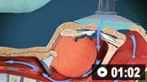
How to use bag-valve-mask apparatus to deliver ventilatory support to adults. Video demonstrates the two-person technique.
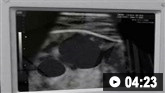
Ultrasound-guided insertion of a non-tunnelled central venous catheter (CVC) into the right internal jugular vein using the Seldinger insertion technique.

How to insert a peripheral venous cannula into the dorsum of the hand.
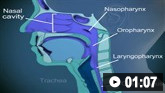
How to select the correct size naspharyngeal airway and insert the airway device safely.
How to size and insert an oropharygeal airway.
Use of this content is subject to our disclaimer