Images and videos
Images


Hip fracture
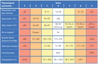
Anteroposterior x-ray showing femoral neck fracture
From the collection of Bradley A. Petrisor, MSc, MD, FRCSC and Mohit Bhandari, MD, MSc, FRCSC
See this image in context in the following section/s:
Hip fracture
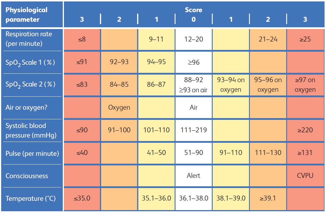
National Early Warning Score 2 (NEWS2) is an early warning score produced by the Royal College of Physicians in the UK. It is based on the assessment of six individual parameters, which are assigned a score of between 0 and 3: respiratory rate, oxygen saturations, temperature, blood pressure, heart rate, and level of consciousness. There are different scales for oxygen saturation levels based on a patient’s physiological target (with scale 2 being used for patients at risk of hypercapnic respiratory failure). The score is then aggregated to give a final total score; the higher the score, the higher the risk of clinical deterioration
Reproduced from: Royal College of Physicians. National Early Warning Score (NEWS) 2: Standardising the assessment of acute-illness severity in the NHS. Updated report of a working party. London: RCP, 2017.
See this image in context in the following section/s:
Hip fracture
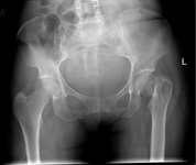
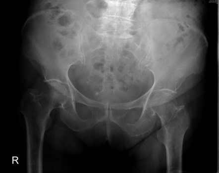
Anteroposterior pelvic radiograph showing a possible intracapsular fracture of the left hip
From the collection of Bradley A. Petrisor, MSc, MD, FRCSC and Mohit Bhandari, MD, MSc, FRCSC
See this image in context in the following section/s:

Hip fracture
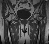
Magnetic resonance imaging showing coronal imaging confirming an intracapsular fracture of the left hip
From the collection of Bradley A. Petrisor, MSc, MD, FRCSC and Mohit Bhandari, MD, MSc, FRCSC
See this image in context in the following section/s:
Hip fracture
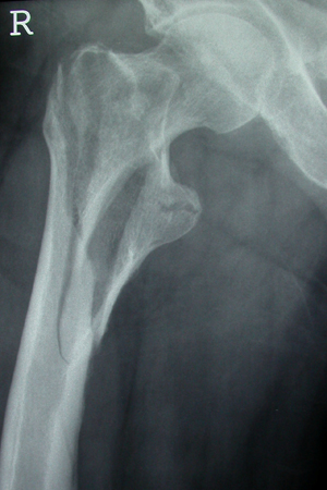
Unstable intertrochanteric fracture on x-ray
From the collection of Bradley A. Petrisor, MSc, MD, FRCSC and Mohit Bhandari, MD, MSc, FRCSC
See this image in context in the following section/s:
Hip fracture
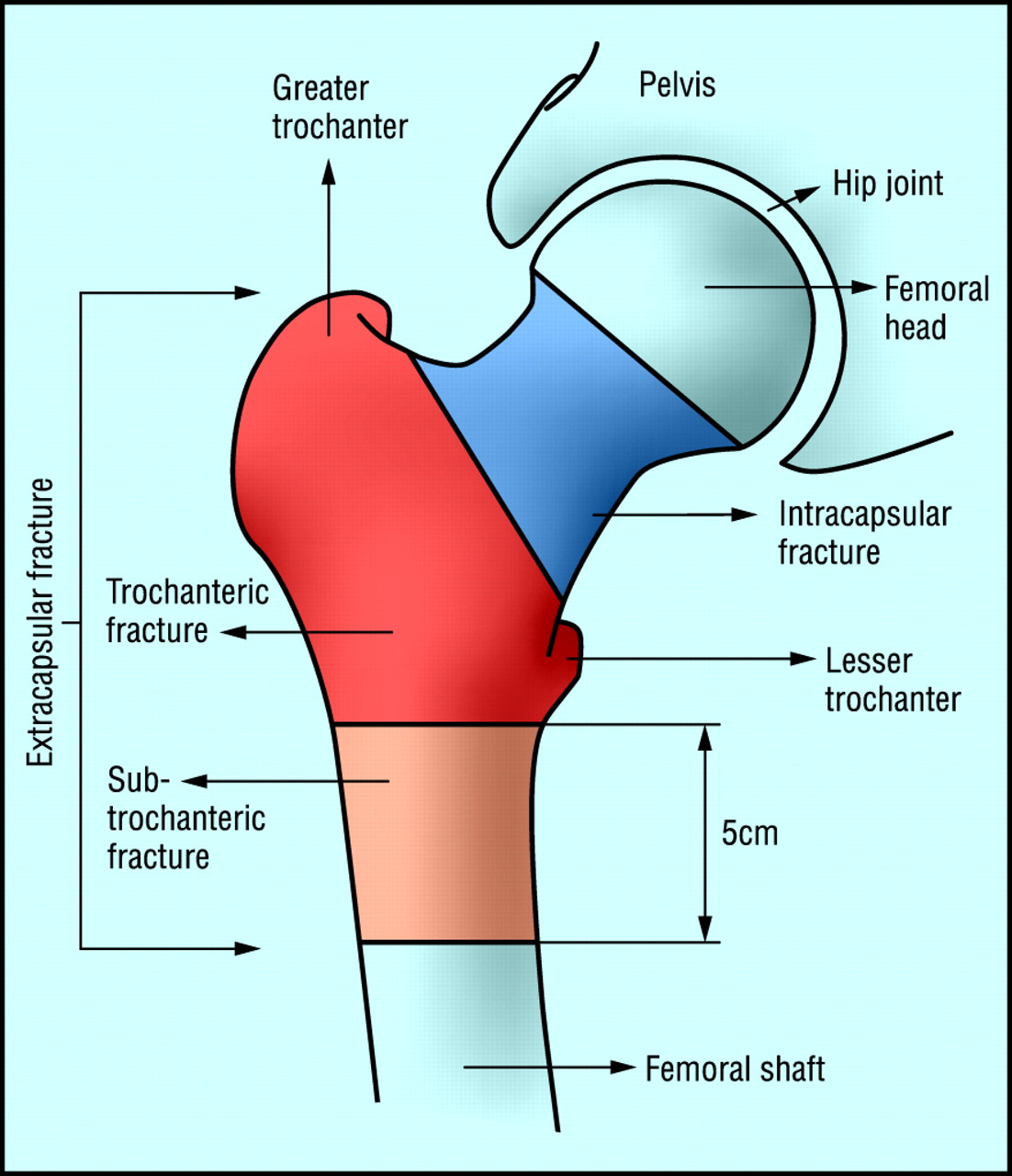
Classification of hip fractures. Fractures in the blue area are intracapsular and those in the red and orange areas are extracapsular
BMJ. 2006 Jul 1;333(7557):27-30
See this image in context in the following section/s:

Hip fracture
Initial anteroposterior radiograph showing a displaced left hip intracapsular fracture
From the collection of Bradley A. Petrisor, MSc, MD, FRCSC and Mohit Bhandari, MD, MSc, FRCSC
See this image in context in the following section/s:

Hip fracture
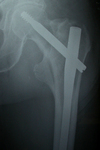
Intramedullary nail (cephalomedullary) for the treatment of an unstable intertrochanteric fracture
From the collection of Bradley A. Petrisor, MSc, MD, FRCSC and Mohit Bhandari, MD, MSc, FRCSC
See this image in context in the following section/s:

Hip fracture
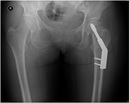
Anteroposterior pelvic radiograph showing a left intracapsular fracture fixed with a sliding hip screw construct
From the collection of Bradley A. Petrisor, MSc, MD, FRCSC and Mohit Bhandari, MD, MSc, FRCSC
See this image in context in the following section/s:
Use of this content is subject to our disclaimer