Article Text

Statistics from Altmetric.com
Description
A 25-year-old man presented to the emergency department reporting a 6-hour history of progressive neck swelling associated with a single episode of chest pain. He had no significant medical history. He initially denied any illicit drug use.
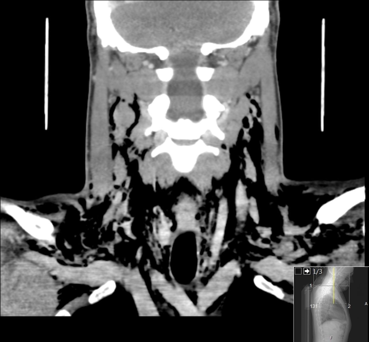
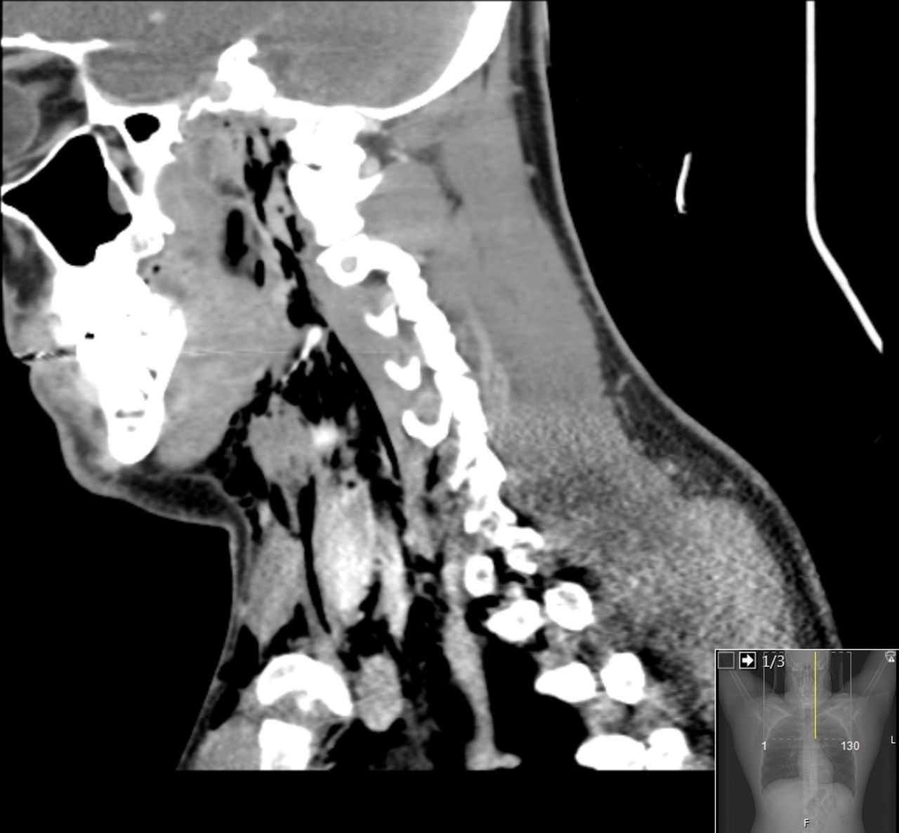
On examination, the neck was visibly distended with palpable crepitus that extended to the upper torso. Cardiorespiratory examination was otherwise unremarkable. He was systemically well and routine observations were normal. The ECG showed widespread concave ST elevation suggestive of pericarditis. The chest radiograph demonstrated pneumomediastinum. A CT scan of the neck and thorax revealed widespread surgical emphysema along the thoracic wall, extending through the mediastinum, upwards into the neck involving both superficial and deep compartments (figure 1) up to the skull base (figure 2). No pneumothorax was seen. The white blood cell count was mildly raised at 17.7×10⁹/L, C reactive protein was 24 mg/L and troponin was negative. Renal and liver function tests were normal.

Coronal CT image demonstrating widespread surgical emphysema extending through superficial and deep fascial planes.

Sagittal CT image demonstrating surgical emphysema extending from level of mediastinum to base of skull.
Differential diagnosis included barotrauma, oesophageal rupture and deep tissue infection.
Given the associated neck swelling, the ENT team were called to review the patient. Flexible nasoendoscopy was performed, which revealed an ulcerated nasal septum but an otherwise normal airway tract. These appearances were strongly suggestive of cocaine abuse. On further enquiry, the patient admitted to snorting cocaine and inhaling 20 nitrous oxide balloons 2 days previously.
The patient was managed with prophylactic antibiotics and close monitoring on the intensive care unit. Symptoms of chest pain and neck swelling resolved within 2 days, and a repeat chest radiograph showed a significant reduction in the pneumomediastinum after 4 days. The patient was discharged with advice to avoid air travel for 3 weeks, in line with the Aerospace Medical Association guidelines for pneumomediastinum.1
Spontaneous (atraumatic) pnuemomediastinum is uncommon, with a reported incidence of between 1:7000 and 1:45 000.2 Inhaled recreational drugs such as cocaine and nitrous oxide have been reported as precipitating factors, along with asthma, emesis and coughing.2 The condition is often self-limiting. Evidence surrounding the use of prophylactic antibiotics is limited, and it is our opinion that this decision should be considered on an individual basis.
Intranasal use of cocaine powder is known to have corrosive effects on respiratory nasal mucosa and smoking cocaine can result in pulmonary complications including pneumomediastinum.3 We postulate that the damaged mucosal integrity coupled with repeated pressurised inhalation and exhalation through a balloon was sufficient to rupture the marginal alveoli, causing air to dissect into the mediastinum and surrounding fascial planes.
Learning points
Spontaneous, atraumatic pneumomediastinum is rare.
A careful history of illicit drug use, including the specific use of cocaine or nitrous oxide, should be sought in all patients presenting with spontaneous pneumomediastinum, as it is an emerging complication of these substances.
Spontaneous pneumomediastinum is often self-limiting, but lifestyle advice should be given to avoid air travel for 3 weeks.
Footnotes
Contributors RR wrote and revised the manuscript, gained patient consent and obtained images. YA wrote and revised the manuscript, and managed the case. MV identified the case, revised the manuscript and managed the case. DG-W revised the manuscript.
Funding The authors have not declared a specific grant for this research from any funding agency in the public, commercial or not-for-profit sectors.
Competing interests None declared.
Patient consent Obtained.
Provenance and peer review Not commissioned; externally peer reviewed.